About the Profundus camera technology
The Profundus camera is an ophthalmic camera, using adaptive optics to image the retina with high resolution for instance. Adaptive optics is a technology of capturing diffraction-limited images in adverse circumstances that would normally lead to strongly degraded image quality and loss of resolution. By implementing Multi Conjugate Adaptive Optics (MCAO), aberrations are measured in different angular directions, and using multiple deformable mirrors it is possible to correct aberrations over a larger Field of View (FOV).
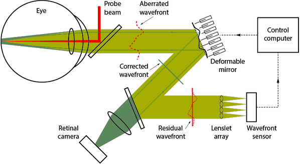
About the general principle of Adaptive Optics (AO) in the eye
A probe beam is focused to a spot on the retina. Reflected light from this retinal beacon will radiate through the optical media (basically the cornea and the lens) and the pupil of the eye. A wavefront sensor is used to measure the aberrations introduced by the optics of the eye, and a control computer is used to analyse the measurements and calculate a correction.
High-resolution imaging of the object is enabled by applying the calculated corrections to a corrective element, usually a deformable mirror, positioned in a pupil conjugate plane in the optical path between the retina and its image on the retinal camera.
Such a system is referred to as a ’single-conjugate AO system’. A multi-conjugate AO system utilizes multiple probe beams and at least two corrective elements to widen the corrected field of view. Retinal features down to a few microns can be resolved by correcting the optical imperfections of the eye.
High-resolution
Up to 10x higher than conventional instruments for retinal imaging
Wide-field
Up to 8x larger field of view compared to devices based on single-conjugate adaptive optics that only use one corrective element
Structural sizes:
Photoreceptors ~ 2-15 µm
Vessels > 5 µm
Resolution
Traditional fundus camera 15-20 µm
Profundus camera 2-3 µm